
Optomap imaging is a crucial part of nearly every eye exam, but what is it really? And how does it work?
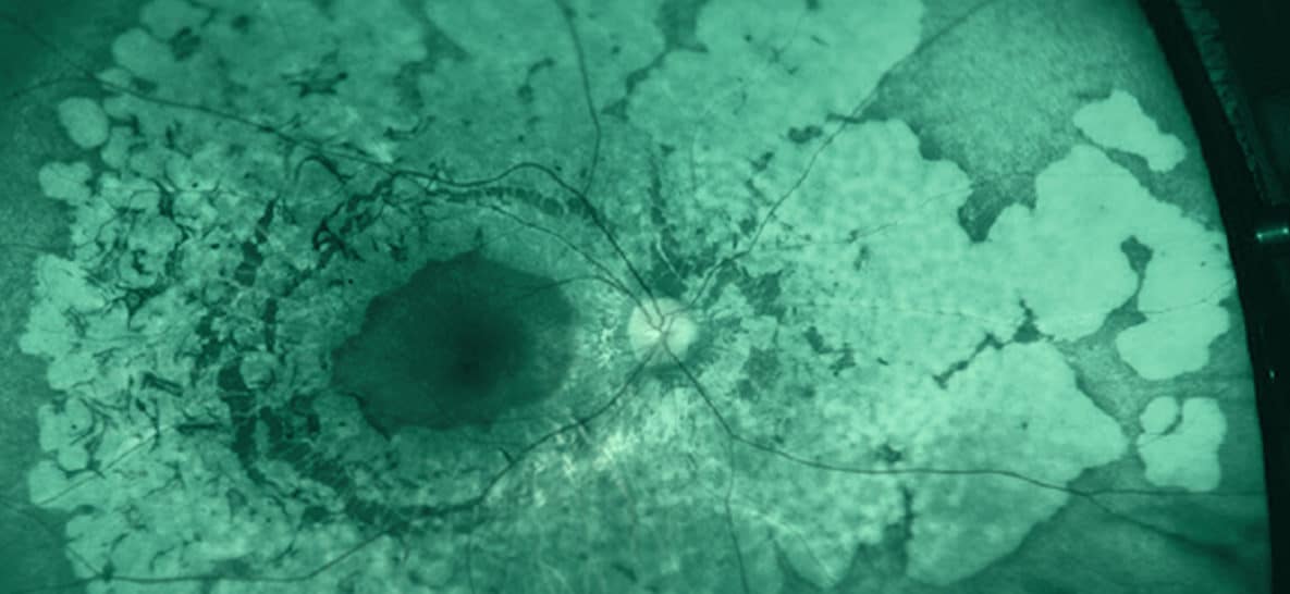
Basically, it entails taking a picture of the retina, which lines the back of the eyeball, and displays it on a computer monitor for a more in-depth look.
The process is as simple as looking into a camera lens, one eye at a time, and holding your eye open for a moment while the image is being captured, generating the most comprehensive view your eye doctor can get. Nothing physically touches your eye at any time.
This image provides a high definition, 200-degree image of your eye, allowing your eye doctor to closely examine the retina and the blood vessels within it. Because of this, eye diseases can be detected earlier, which decreases the chances of lost vision due to these diseases. Because of the massive benefit, Optomap imaging is a routine test at almost all eye exams.
Furthermore, if you are getting Optomap imaging done at every exam, your eye doctor can compare your past images with current ones to ascertain if there are any changes over time.
Diseases of the eye are not the only diseases Optomap imaging can detect. With the close-up view of the retina and its blood vessels, diseases such as diabetes, hypertension, and stroke can be detected long before they manifest any other signs and symptoms. As with any other diseases, the earlier detection the better. It is much easier to intervene and treat a disease in its early stages than it is to try to fix the damage that has already been done by not catching and diagnosing a problem sooner.
If your eye doctor does not provide Optomap imaging, consider requesting to have it done. With it being more effective and efficient than a traditional retinal exam, you will be doing both your sight and your overall health a favor.
Comments are closed.
